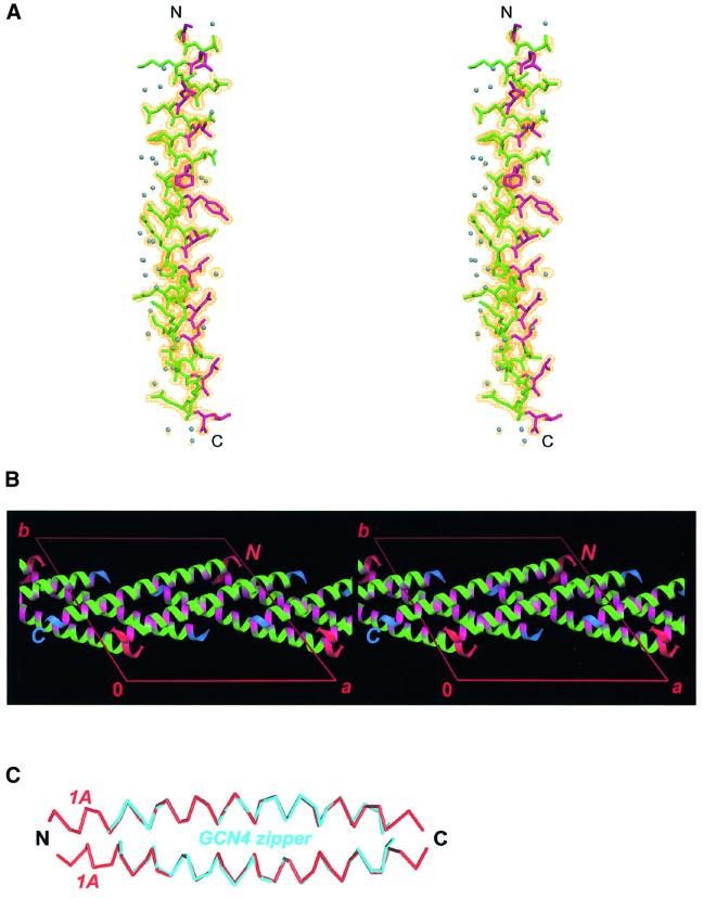
Fig. 2. Crystal structure of the vimentin fragment 1A. (A) Stereo view of the atomic model and electron density map with coefficients 2Fobs – Fcalc contoured at 1.2σ. Residues in the a and d positions of the putative heptad repeat are shown in magenta. Solvent molecules are shown as blue spheres. (B) The crystal packing arrangement of 1A shown in stereo. The a and d positions are highlighted with magenta. The N- and C-termini of the helices are marked in red and blue, respectively. (C) Modeling of a parallel coiled-coil by docking two 1A helices (red) while using the GCN4 zipper structure (cyan) as a ruler.

An official website of the United States government
Here's how you know
Official websites use .gov
A
.gov website belongs to an official
government organization in the United States.
Secure .gov websites use HTTPS
A lock (
) or https:// means you've safely
connected to the .gov website. Share sensitive
information only on official, secure websites.
