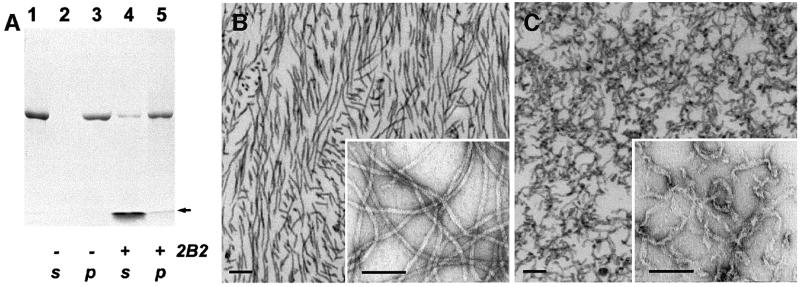
Fig. 6. Effect of the 2B2 fragment on IF assembly. (A) Denaturing gel electrophoresis (SDS–PAGE) illustrating the assembly of human recombinant vimentin alone (lanes 2 and 3) and in the presence of the 2B2 fragment (lanes 4 and 5). The 2B2 fragment was added to the vimentin sample (lane 1) at a 10-fold molar excess, and then filament assembly in the test and reference (i.e. without the fragment addition) samples was performed as described in Materials and methods. The samples subsequently were centrifuged in a Beckman Airfuge for 30 min at 10 p.s.i. yielding the supernatant (lanes 2 and 4) and pelleted (lanes 3 and 5) fractions. The arrow indicates the location of the gel front. (B) Negatively stained EM images of vimentin IFs assembled in vitro. The samples were prepared by ultrathin sectioning (main figure) or on grids (inset). Scale bars are 100 nm. (C) Similarly assembled IFs, which subsequently were incubated with a 10-fold molar excess of the 2B2 fragment for 1 h at 37°C.

An official website of the United States government
Here's how you know
Official websites use .gov
A
.gov website belongs to an official
government organization in the United States.
Secure .gov websites use HTTPS
A lock (
) or https:// means you've safely
connected to the .gov website. Share sensitive
information only on official, secure websites.
