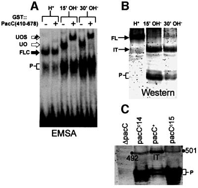
Fig. 3. The transition of the ‘closed’ to the ‘open’ PacC forms strictly correlates with the formation of a new PacC polypeptide. (A) Wild-type cells grown under acidic conditions (H+) were shifted to alkaline conditions (OH–) and protein extracts from the indicated time points were analysed by EMSA. The ‘open’ PacC form was revealed by the supershift assay with GST::PacC(410–678). Abbreviations are as in Figure 2. (B) Western analysis of extracts used in (A) with α-PacC(5–265) antiserum. FL, full-length PacC; IT, intermediate; P, processed form. (C) The C-terminal residue of the intermediate lies between residues 493 and 500. The primary translation products of two truncating pacCc alleles (leading to signal-independent processing) ending at residues 492 and 501, respectively, were used as internal standards to calibrate the size of the intermediate in a western blot where proteins were detected with α-PacC(5–265) antiserum. The size of the intermediate is clearly >492 residues and <501 residues.

An official website of the United States government
Here's how you know
Official websites use .gov
A
.gov website belongs to an official
government organization in the United States.
Secure .gov websites use HTTPS
A lock (
) or https:// means you've safely
connected to the .gov website. Share sensitive
information only on official, secure websites.
