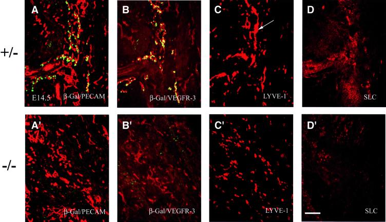
Fig. 3. At E14.5, widespread expression of different lymphatic markers in Prox1 heterozygous embryos indicates the progression of lymphangiogenesis. Panels correspond to transverse sections taken at the level of the forelimb. (A) The lymphatic vessel network, which is indicated by the co-expression of β-gal/Prox1 (green) and CD31 (red), is interspersed with the blood vessels and capillaries, which are indicated by the expression of CD31 alone. The patterns of expression of VEGFR-3 (B), LYVE-1 (C) and SLC (D) overlap with that of Prox1/β-gal. LYVE-1 expression is detected in capillary-like structures (C, arrow) and in some scattered cells. (A′–D′) The absence of lymphatic vasculature in Prox1 nullizygous embryos is corroborated by the absence of VEGFR-3, SLC and LYVE-1 expression in endothelial cells at E14.5. (A′) CD31 expression in Prox1 null embryos indicates that the formation of the blood vascular system is normal in these mice. (B′) Low levels of VEGFR-3 expression are detected in only the blood vascular system. (C′) LYVE-1 expression is present, but only in scattered macrophages. (D′) No endothelial-specific expression of SLC is detected. Scale bar: 100 µm.

An official website of the United States government
Here's how you know
Official websites use .gov
A
.gov website belongs to an official
government organization in the United States.
Secure .gov websites use HTTPS
A lock (
) or https:// means you've safely
connected to the .gov website. Share sensitive
information only on official, secure websites.
