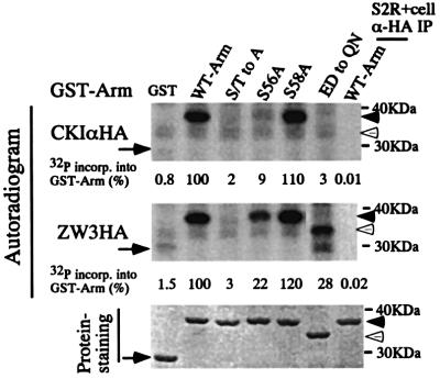
Fig. 9. In vitro kinase experiment to determine the major phosphorylation site for CKIα and ZW3 in the N-terminal region of Arm. Various GST–Arm fusion proteins were phosphorylated by CKIα-HA (upper panel) and ZW3-HA (middle panel). HA-immunoprecipitate from naive S2R+ cells was used as a negative control (the right lane in each panel). The same amount (10 µg) of GST or GST–Arm fusion proteins were used for each reaction (50 µl) and reaction mixtures were incubated at 30°C for 10 min, before 10 µl of each was subjected to SDS–PAGE. The bottom panel shows the staining profiles of GST–Arm fusion proteins in the dried gel, from which the autoradiogram shown in the top panel was generated. The arrows and the arrowheads indicate the migration positions of plain GST and GST–Arm fusion proteins, respectively. The open arrow heads show the migration positions of a GST–Arm protein with the ED to QN mutation, which has a higher electrophoretic mobility. The amount of 32P incorporated into each GST–Arm fusion protein was expressed as a percentage of the amount incorporated into the protein containing the wild-type Arm sequence.

An official website of the United States government
Here's how you know
Official websites use .gov
A
.gov website belongs to an official
government organization in the United States.
Secure .gov websites use HTTPS
A lock (
) or https:// means you've safely
connected to the .gov website. Share sensitive
information only on official, secure websites.
