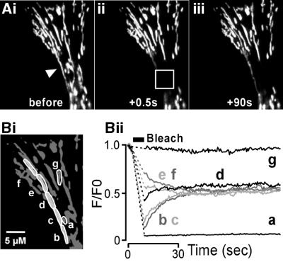
Fig. 3. DsRed1 is rapidly diffusible in the mitochondrial matrix. (Ai) A portion of a HeLa cell expressing mito-DsRed1. Both long (denoted by an arrow) and short discrete mitochondria can be seen in this region. The region bounded by the white box in (Aii) was photobleached using a 5 s illumination as described in Materials and methods. The bleached region encompassed the middle portion of the long mitochondria and completely surrounded several smaller mitochondria. The images in (Aii) and (Aiii) show the gradual recovery of fluorescence in the long mitochondrion, and were obtained at times corresponding to 0.5 and 90 s after the photobleach. (B) A quantitation of the fluorescence recovery within the long mitochondrion. The fluorescence was monitored in the regions denoted in (Bi). Areas a, b, c and d were within the bleached zone, but e, f and g were not. The traces in (Bii) illustrate the intensity of mito-DsRed1 fluorescence in these regions before and after the photobleach. Note that regions a and g were not part of the long mitochondrion. Scale bar, 5 µm. The data presented show a typical response observed in more than five cells.

An official website of the United States government
Here's how you know
Official websites use .gov
A
.gov website belongs to an official
government organization in the United States.
Secure .gov websites use HTTPS
A lock (
) or https:// means you've safely
connected to the .gov website. Share sensitive
information only on official, secure websites.
