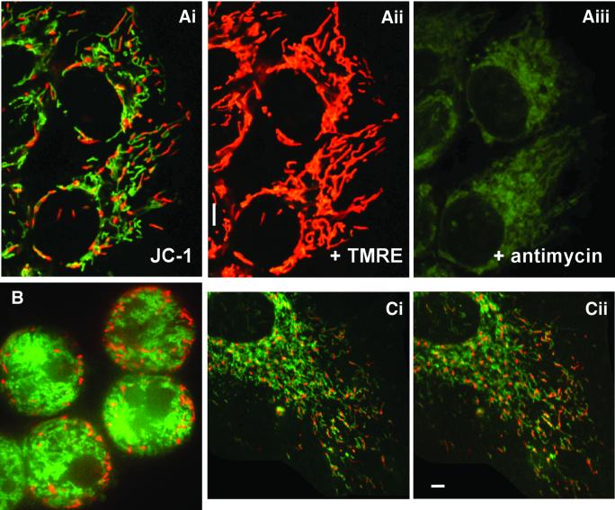
Fig. 5. Heterogeneity in membrane potential revealed by JC-1. (Ai) HeLa cell mitochondria after incubation with JC-1, illustrating the heterogeneity in mitochondria membrane potential within a single cell. To demonstrate co-localization of TMRE with JC-1, the same cells were subsequently co-loaded with TMRE (0.1 µM, 20 min). As illustrated in (Aii), the same organelles are stained. The mitochondria within the cells were then depolarized by addition of antimycin (10 µM) plus oligomycin (2 µM). (B) The peripheral location of red staining mitochondria in a hepatocyte stained with JC-1 at 22°C. (Ci and ii) A section of the same JC-1-loaded HUVEC cell at 22 and 37°C. The HUVEC cell was incubated in JC-1 at 22°C for 1 h before the image in (Ci) was obtained. The cell was then slowly warmed to 37°C (∼10 min) and then incubated for a further 30 min in the continual presence of JC-1. Scale bar, 5 µm.

An official website of the United States government
Here's how you know
Official websites use .gov
A
.gov website belongs to an official
government organization in the United States.
Secure .gov websites use HTTPS
A lock (
) or https:// means you've safely
connected to the .gov website. Share sensitive
information only on official, secure websites.
