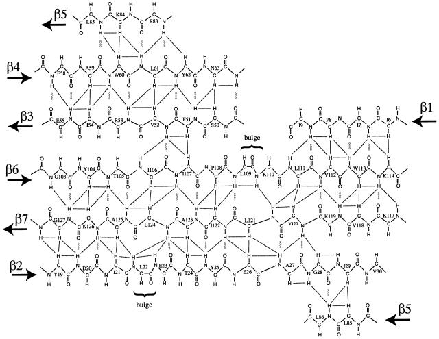
Fig. 5. β-sheet structure of the SmpB protein. Note that strand β5 appears twice in the figure, to show its anti-parallel arrangement relative to strands β2 and β4, completing the closed β-barrel structure. Some of the pairs of protons for which NOE cross peaks are observed are indicated by lines. Inter-strand hydrogen bonds are indicated by dotted lines.

An official website of the United States government
Here's how you know
Official websites use .gov
A
.gov website belongs to an official
government organization in the United States.
Secure .gov websites use HTTPS
A lock (
) or https:// means you've safely
connected to the .gov website. Share sensitive
information only on official, secure websites.
