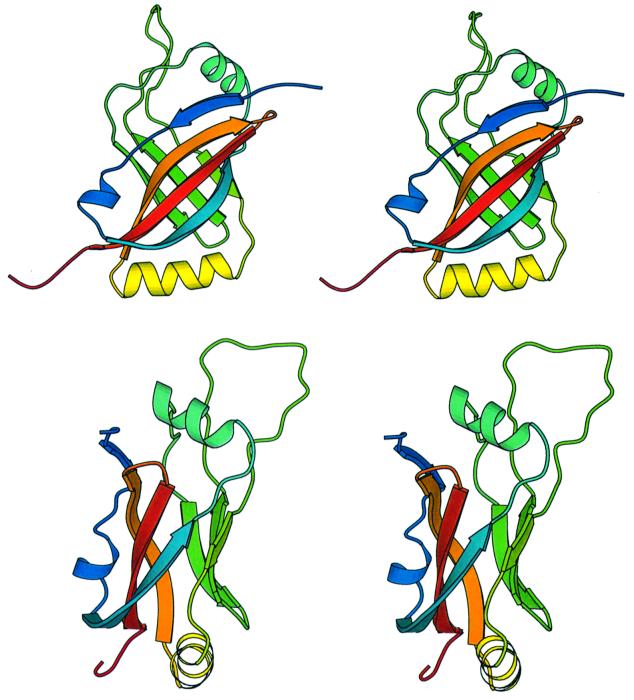
Fig. 6. Stereoview ribbon diagrams of SmpB. The two views differ by a 90° rotation. The protein is color-ramped from blue at the N-terminus to red at the C-terminus. The coordinates for SmpB have been submitted to the Protein Data Bank and have been assigned PDB code 1K8H. The diagram was created using MOLSCRIPT (Kraulis, 1991).

An official website of the United States government
Here's how you know
Official websites use .gov
A
.gov website belongs to an official
government organization in the United States.
Secure .gov websites use HTTPS
A lock (
) or https:// means you've safely
connected to the .gov website. Share sensitive
information only on official, secure websites.
