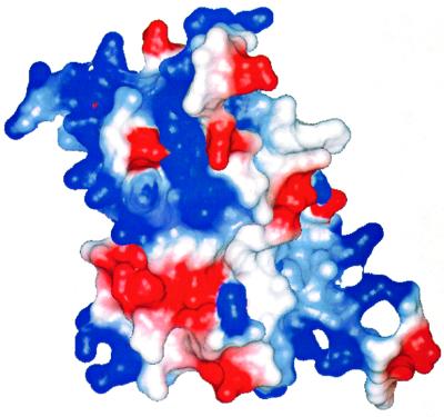
Fig. 8. Electrostatic surface potential plot for SmpB. The view faces strands β3, β4 and β5, where the majority of the conserved surface amino acids are visible. The surface charge is predominantly positive (blue) on the surfaces containing the highest density of conserved residues (upper right and lower left), indicating regions of likely interaction with RNA. It is noted, however, that negatively charged amino acids (red) can also make specific interacts with RNA bases by serving as hydrogen bond acceptors. The figure was prepared using the program MOLMOL (Koradi et al., 1996).

An official website of the United States government
Here's how you know
Official websites use .gov
A
.gov website belongs to an official
government organization in the United States.
Secure .gov websites use HTTPS
A lock (
) or https:// means you've safely
connected to the .gov website. Share sensitive
information only on official, secure websites.
