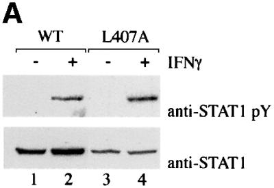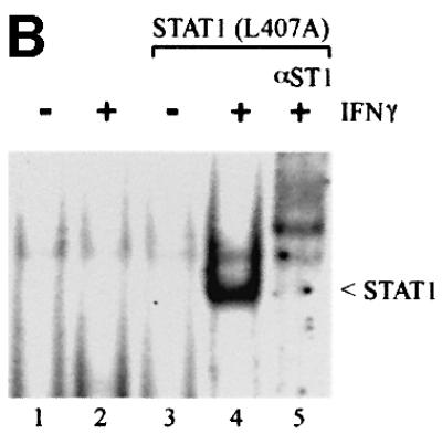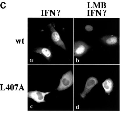


Fig. 2. STAT1–GFP(L407A) is tyrosine phosphorylated and binds DNA but does not translocate to the nucleus. (A) U3A cells expressing STAT1–GFP (lanes 1 and 2) or STAT1–GFP(L407A) (lanes 3 and 4) were untreated (–) or treated with IFN-γ for 30 min (+). Proteins from cell lysates were immunoprecipitated with anti-STAT1 antibody and western blots were performed with anti-STAT1 phosphotyrosine antibody (anti-STAT1 pY) (upper panel) or anti-STAT1 antibody (lower panel). (B) EMSA was performed with the IRF-1 GAS probe and whole-cell lysates from U3A cells (lanes 1 and 2) or U3A cells expressing STAT1–GFP (L407A) (lanes 3–5) were either untreated (–) or treated with IFN-γ for 30 min (+). Anti-STAT1 antibody (lane 5) was added to the DNA binding reaction. (C) U3A cells expressing wtSTAT1–GFP or STAT1–GFP(L407A) were treated with IFN-γ for 30 min, with or without pre-treatment with LMB. Cellular localization was examined by fluorescent microscopy.
