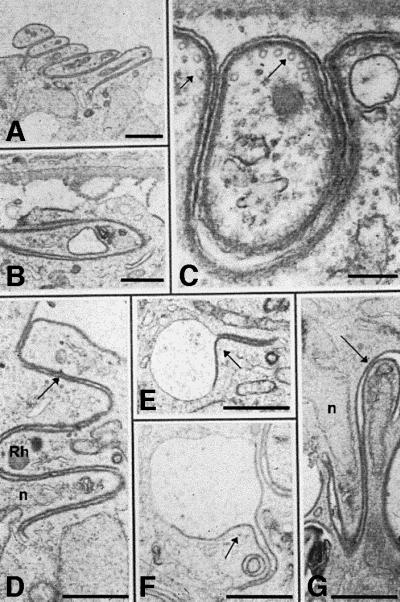
Fig. 5. Electron micrographs of CSko oocysts showing aberrant budding in the subcapsular space (A–C) and in the sporoblast cytoplasm (D–G). (A) Parallel stacking of bud-like structures. Bar: 3 µm. (B) An abnormal bud extends parallel to the sporoblast periphery instead of away into the subcapsular space. Bar: 2.5 µm. (C) Inner membranes and microtubules (arrows) underneath the oocyst plasma membrane and the bud outer membrane. Bar: 0.5 µm. (D–G) Cytoplasmic membranes lined with inner membranes (arrows), either totally (D and G) or partially (E and F). Rh, rhoptry; n, nucleus. Bars: 2.5 µm.
