Abstract
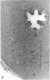
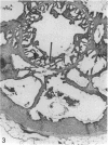
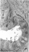
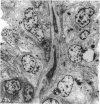
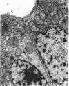
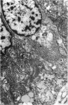
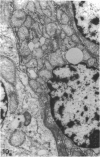
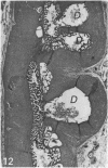
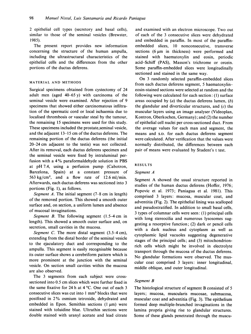
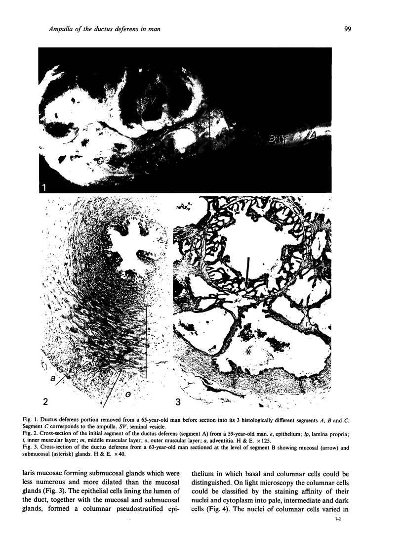
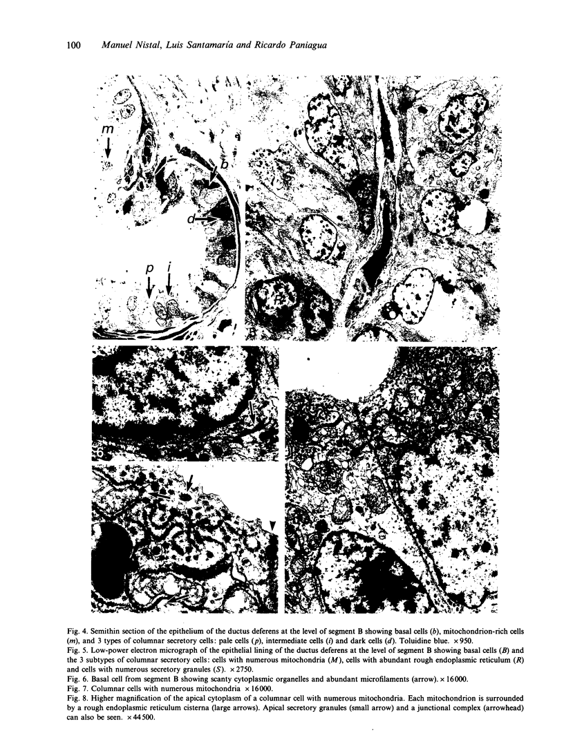
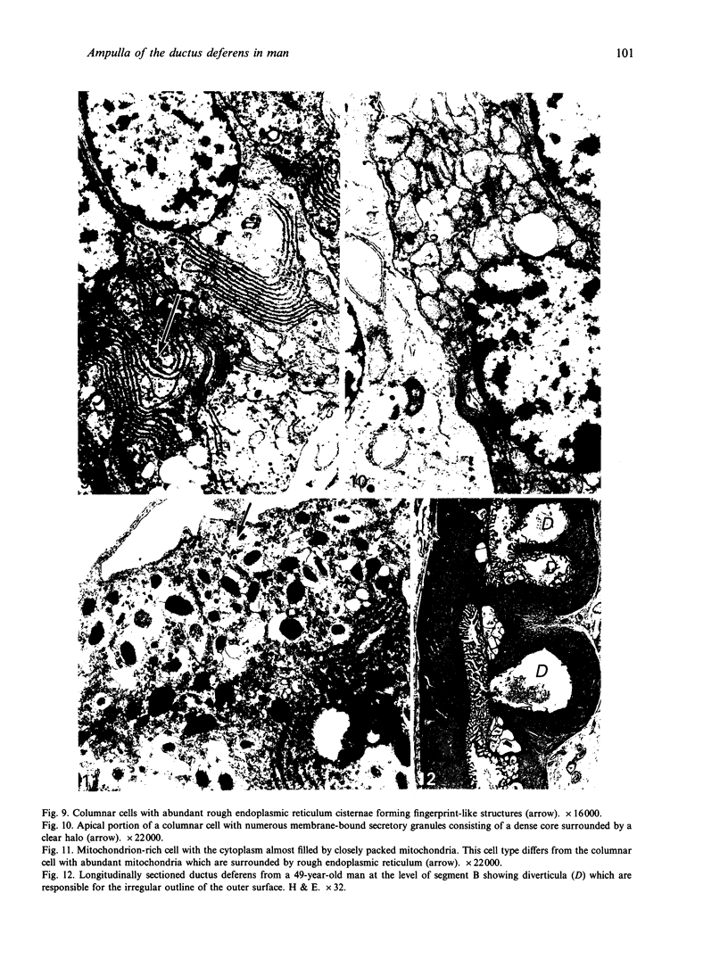
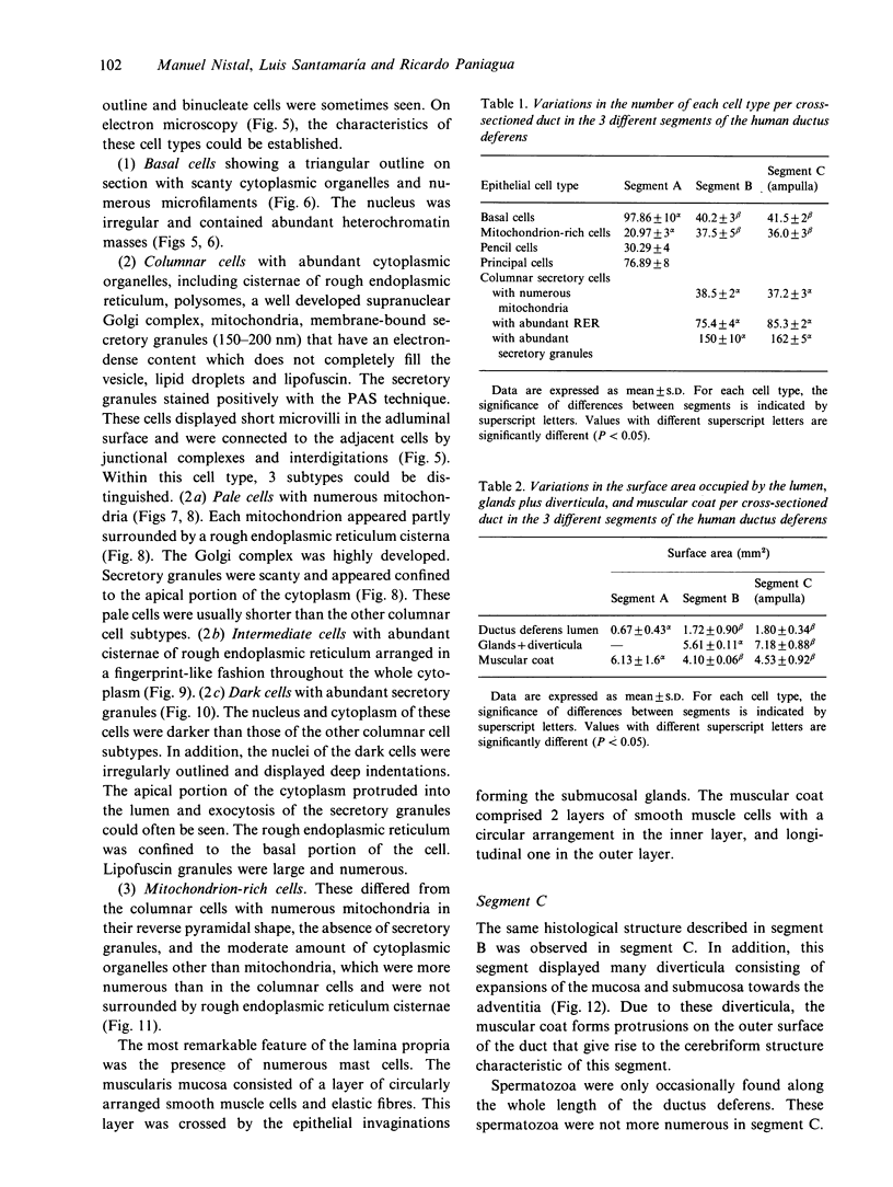
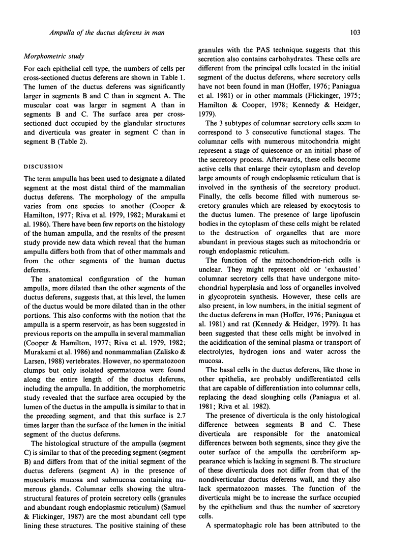
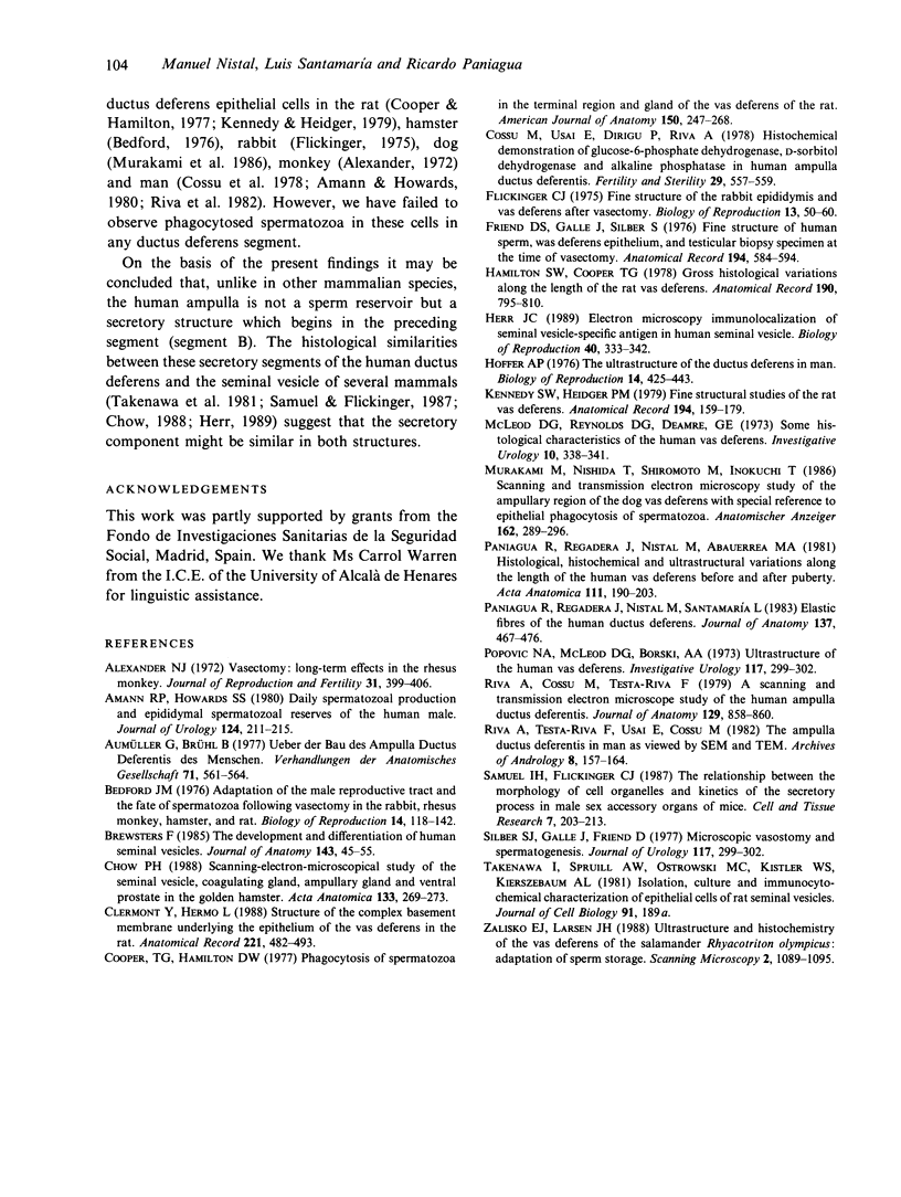
In order to compare the histology of the ampulla of the ductus deferens with that of the other segments of the duct in man, the seminal vesicle and the adjacent 13-15 cm of the ductus deferens were obtained during cystectomy from 15 adult men, and were processed for light and electron microscopy. Each ductus deferens specimen was divided into 3 segments: segment A or initial segment (the most proximal to the testis) showing a smooth outer surface and, on section, a uniform lumen and absence of mucosal invaginations; segment B (1.5-4 cm) showing a smooth outer surface and, on section, small cavities in the mucosa; and segment C or ampulla (3-4 cm), which was easily recognisable because of the cerebriform pattern on its outer surface. Segment A showed the usual histological pattern reported in studies of the human ductus deferens. Segment B consisted of mucosa, muscularis mucosae, submucosa, muscular coat and adventitia. The epithelial lining formed multiple branched invaginations in the lamina propria and submucosa giving rise to glandular structures. The lumen of the duct and the glands were lined by the same cell types: (1) basal cells; (2) mitochondrion-rich cells; and (3) columnar cells with the ultrastructural features of glycoprotein-secreting cells. The latter cells could be classified into 3 subtypes suggesting different stages of development: (a) with abundant mitochondria; (b) with abundant rough endoplasmic reticulum; and (c) with abundant secretory granules. Segment C or the ampulla showed the same histology as segment B except for the presence of many diverticula in the ampulla.(ABSTRACT TRUNCATED AT 250 WORDS)
Full text
PDF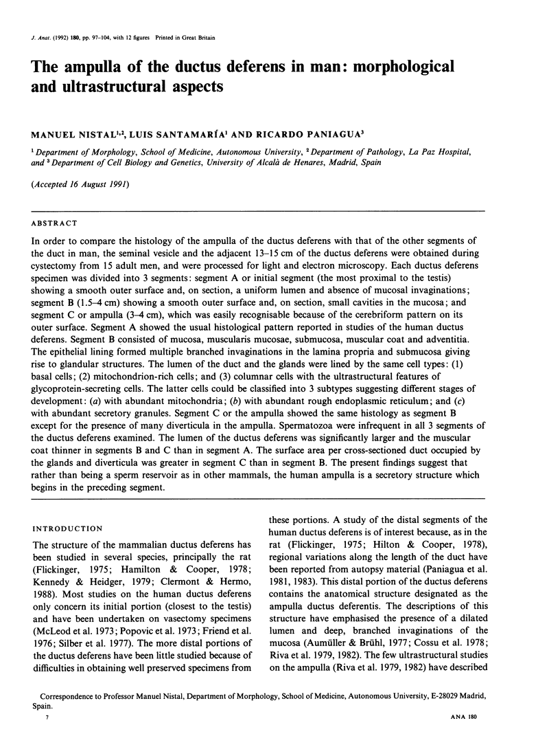
Images in this article
Selected References
These references are in PubMed. This may not be the complete list of references from this article.
- Alexander N. J. Vasectomy: long-term effects in the rhesus monkey. J Reprod Fertil. 1972 Dec;31(3):399–406. doi: 10.1530/jrf.0.0310399. [DOI] [PubMed] [Google Scholar]
- Amann R. P., Howards S. S. Daily spermatozoal production and epididymal spermatozoal reserves of the human male. J Urol. 1980 Aug;124(2):211–215. doi: 10.1016/s0022-5347(17)55377-x. [DOI] [PubMed] [Google Scholar]
- Aumüller G., Brühl B. Uber den Bau der Ampulla ductus deferentis des Menschen. Verh Anat Ges. 1977;(71 Pt 1):561–564. [PubMed] [Google Scholar]
- Bedford J. M. Adaptations of the male reproductive tract and the fate of spermatozoa following vasectomy in the rabbit, rhesus monkey, hamster and rat. Biol Reprod. 1976 Mar;14(2):118–142. doi: 10.1095/biolreprod14.2.118. [DOI] [PubMed] [Google Scholar]
- Brewster S. F. The development and differentiation of human seminal vesicles. J Anat. 1985 Dec;143:45–55. [PMC free article] [PubMed] [Google Scholar]
- Chow P. H. Scanning-electron-microscopical study of the seminal vesicle, coagulating gland, ampullary gland and ventral prostate in the golden hamster. Acta Anat (Basel) 1988;133(4):269–273. doi: 10.1159/000146653. [DOI] [PubMed] [Google Scholar]
- Clermont Y., Hermo L. Structure of the complex basement membrane underlying the epithelium of the vas deferens in the rat. Anat Rec. 1988 May;221(1):482–493. doi: 10.1002/ar.1092210105. [DOI] [PubMed] [Google Scholar]
- Cooper T. G., Hamilton D. W. Phagocytosis of spermatozoa in the terminal region and gland of the vas deferens of the rat. Am J Anat. 1977 Oct;150(2):247–267. doi: 10.1002/aja.1001500204. [DOI] [PubMed] [Google Scholar]
- Cossu M., Usai E., Sirigu P., Riva A. Histochemical demonstration of glucose-6-phosphate dehydrogenase, D-sorbitol dehydrogenase, and alkaline phosphatase in human ampulla ductus deferentis. Fertil Steril. 1978 May;29(5):557–559. doi: 10.1016/s0015-0282(16)43285-1. [DOI] [PubMed] [Google Scholar]
- Flickinger C. J. Fine structure of the rabbit epididymis and vas deferens after vasectomy. Biol Reprod. 1976 Aug;13(1):50–60. doi: 10.1095/biolreprod13.1.50. [DOI] [PubMed] [Google Scholar]
- Hamilton D. W., Cooper T. G. Gross and histological variations along the length of the rat vas deferens. Anat Rec. 1978 Apr;190(4):795–809. doi: 10.1002/ar.1091900403. [DOI] [PubMed] [Google Scholar]
- Herr J. C., Spell D. R., Conklin D. J., Flickinger C. J. Electron microscopic immunolocalization of seminal vesicle-specific antigen in human seminal vesicle. Biol Reprod. 1989 Feb;40(2):333–342. doi: 10.1095/biolreprod40.2.333. [DOI] [PubMed] [Google Scholar]
- Hoffer A. P. The ultrastructure of the ductus deferens in man. Biol Reprod. 1976 May;14(4):425–443. doi: 10.1095/biolreprod14.4.425. [DOI] [PubMed] [Google Scholar]
- Kennedy S. W., Heidger P. M., Jr Fine structural studies of the rat vas deferens. Anat Rec. 1979 May;194(1):159–179. doi: 10.1002/ar.1091940111. [DOI] [PubMed] [Google Scholar]
- McLeod D. G., Reynolds D. G., Demaree G. E. Some pharmacologic characteristics of the human vas deferens. Invest Ophthalmol. 1973 Mar;10(5):338–341. [PubMed] [Google Scholar]
- Murakami M., Nishida T., Shiromoto M., Inokuchi T. Scanning and transmission electron microscopic study of the ampullary region of the dog vas deferens, with special reference to epithelial phagocytosis of spermatozoa and latex beads. Anat Anz. 1986;162(4):289–296. [PubMed] [Google Scholar]
- Paniagua R., Regadera J., Nistal M., Abaurrea M. A. Histological, histochemical and ultrastructural variations along the length of the human vas deferens before and after puberty. Acta Anat (Basel) 1982;111(3):190–203. doi: 10.1159/000145467. [DOI] [PubMed] [Google Scholar]
- Paniagua R., Regadera J., Nistal M., Santamaría L. Elastic fibres of the human ductus deferens. J Anat. 1983 Oct;137(Pt 3):467–476. [PMC free article] [PubMed] [Google Scholar]
- Riva A., Testa-Riva F., Usai E., Cossu M. The ampulla ductus deferentis in man, as viewed by SEM and TEM. Arch Androl. 1982 May;8(3):157–164. doi: 10.3109/01485018208987034. [DOI] [PubMed] [Google Scholar]
- Samuel L. H., Flickinger C. J. The relationship between the morphology of cell organelles and kinetics of the secretory process in male sex accessory glands of mice. Cell Tissue Res. 1987 Jan;247(1):203–213. doi: 10.1007/BF00216563. [DOI] [PubMed] [Google Scholar]
- Silber S. J., Galle J., Friend D. Microscopic vasovasostomy and spermatogenesis. J Urol. 1977 Mar;117(3):299–302. doi: 10.1016/s0022-5347(17)58440-2. [DOI] [PubMed] [Google Scholar]
- Zalisko E. J., Larsen J. H., Jr Ultrastructure and histochemistry of the vas deferens of the salamander Rhyacotriton olympicus: adaptations for sperm storage. Scanning Microsc. 1988 Jun;2(2):1089–1095. [PubMed] [Google Scholar]


