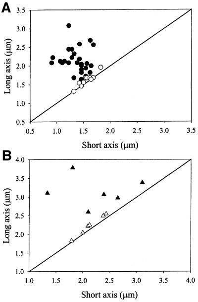
Fig. 5. Patterns of GFP–MinD and MinE–GFP movement correlate with cell geometry. (A) A total of 830 individual movements of GFP–MinD in 40 cells of WM1534 were monitored by time-lapse microscopy in fields similar to those shown in Figure 4, with a range of 14–41 movements per cell. Based on the measured dimensions of each of the 40 cells, the GFP–MinD movements were defined as either along the long axis, along the short axis or along multiple axes. Twenty-nine cells were defined as having predominantly long-axis migrations (filled circles), and the remaining 11 cells had movement to multiple sites (open circles). Of the 29 cells with regular long-axis migrations, three also exhibited occasional migrations along the short axis; this occurred twice out of 27 total migrations, once out of 26 total migrations and twice out of 38 total migrations, respectively, for each of these three cells. (B) A total of 29 MinE–GFP movements were measured in six cells with obvious long axes; all 29 movements were along the long axis (filled triangles). Another six cells were judged to have movement of MinE–GFP in multiple directions (open triangles). In both (A) and (B), the type of movement for each cell in the graph is shown superimposed upon the cell dimensions, with the diagonal line representing a perfectly round cell.

An official website of the United States government
Here's how you know
Official websites use .gov
A
.gov website belongs to an official
government organization in the United States.
Secure .gov websites use HTTPS
A lock (
) or https:// means you've safely
connected to the .gov website. Share sensitive
information only on official, secure websites.
