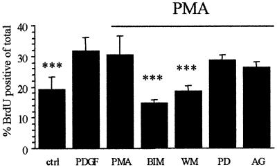
Fig. 5. Effect of PKC activation on the proliferative capacity of OPs on Vn. OPs were either left untreated (ctrl, PDGF, PMA) or were pre- exposed to PD098059 (PD, 50 µM), AG1295 (AG, 10 µM), wortmannin (WM, 50 nM) or BIM (0.5 µM) for 30 min at 37°C (in suspension), plated on Vn (10 µg/ml) and subsequently left untreated (ctrl) or treated with either 1 ng/ml PDGF (PDGF) or 100 nM PMA (all others, indicated by the horizontal bar), and the effect on proliferation was determined as described in Materials and methods. Values shown are means ± SD of at least three independent experiments, each in duplicate. Statistical significance is shown (***P <0.001) between PMA and the other indicated conditions. Note that PKC activation (in the absence of PDGF) via PMA mimicked the PDGF-mediated enhanced proliferation on Vn, and that inhibition of both PKC (BIM) and PI3K (WM) was able to abolish this enhanced proliferation.

An official website of the United States government
Here's how you know
Official websites use .gov
A
.gov website belongs to an official
government organization in the United States.
Secure .gov websites use HTTPS
A lock (
) or https:// means you've safely
connected to the .gov website. Share sensitive
information only on official, secure websites.
