

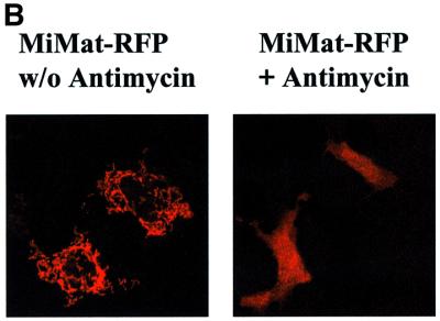
Fig. 3. Import of PorBIA does not depend on an intact mitochondrial membrane potential. (A and B) HeLa cells were transfected with constructs encoding the indicated proteins, stained with Mitotracker, fixed and co-stained with antibodies against the FLAG (PorBIA, VDAC) or Myc (Tom20) tags. Where indicated, the cells were treated with the uncoupling agent antimycin at 100 µM prior to transfection. Both single colours and overlays are shown of confocal sections. Note that in the case of MiMat–RFP, no double staining with Mitotracker could be performed, as both dyes emit light at similar wavelengths.