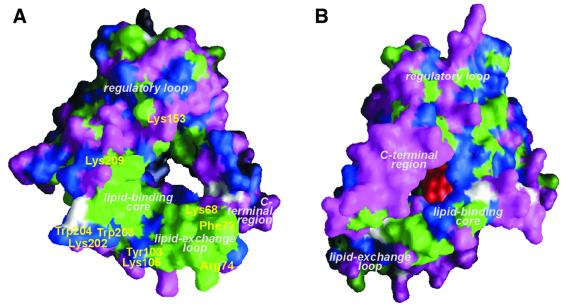
Fig. 2. Molecular surface representation of apo-PITPα showing the openings of the lipid-binding cavity. (A) Viewed from the side of membrane association showing exposed hydrophobic residues (green), charged residues (magenta), polar residues (blue) and glycine (white). The positions of residues thought to be involved in binding to the lipid layer at the interfacial region are indicated in yellow. (B) Viewed from the opposite side (using the same color code) with phosphorylinositol shown at its putative binding site in red.

An official website of the United States government
Here's how you know
Official websites use .gov
A
.gov website belongs to an official
government organization in the United States.
Secure .gov websites use HTTPS
A lock (
) or https:// means you've safely
connected to the .gov website. Share sensitive
information only on official, secure websites.
