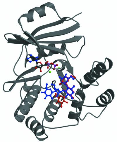
Fig. 1. Crystal structures of APH(3′)-IIIa in complex with ADP and kanamycin A or neomycin B. Ribbon representation of the APH(3′)-IIIa ternary complexes showing the location of the antibiotic binding site. Kanamycin (red) and neomycin (blue) are superimposed in the binding site. Magnesium ions are shown in green. Since the protein structure does not differ significantly between the two ternary complexes, only one is shown.

An official website of the United States government
Here's how you know
Official websites use .gov
A
.gov website belongs to an official
government organization in the United States.
Secure .gov websites use HTTPS
A lock (
) or https:// means you've safely
connected to the .gov website. Share sensitive
information only on official, secure websites.
