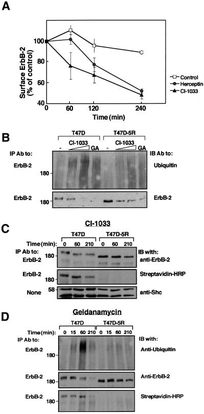
Fig. 3. CI-1033 and GA enhance destruction of both mature and immature ErbB-2 molecules. (A) Monolayers of T47D cells were incubated at 37°C for the indicated time intervals in the presence of either CI-1033 (10 µM; triangles) or Herceptin (10 µg/ml; circles). Control monolayers were incubated in the absence of agents (squares). Levels of cell surface ErbB-2 were determined by incubation with a radiolabeled antibody. Each point represents the average ± SD of duplicates. (B) T47D or T47D-5R cells were treated for 3 h with increasing concentrations of CI-1033 (2 or 20 µM) or with GA (1 µM). Cell extracts were subjected to IP and IB with the indicated antibodies. Note the faster mobility of the nascent ErbB-2 of T47D-5R cells. (C) Cells were treated with CI-1033 (5 µM) for the indicated time intervals, followed by surface labeling for 40 min on ice with Biotin-X-NHS. Streptavidin–HRP denotes incubation of the membrane with streptavidin conjugated to peroxidase. (D) T47D or T47D-5R cells were treated with GA (1 µM) for the indicated time intervals, followed by surface labeling for 40 min on ice with Biotin-X-NHS. Cell extracts were analyzed with the indicated antibodies.

An official website of the United States government
Here's how you know
Official websites use .gov
A
.gov website belongs to an official
government organization in the United States.
Secure .gov websites use HTTPS
A lock (
) or https:// means you've safely
connected to the .gov website. Share sensitive
information only on official, secure websites.
