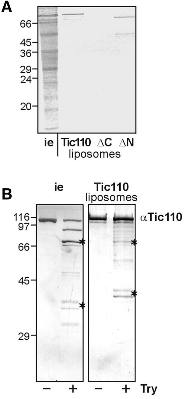
Fig. 2. Reconstitution of Tic110 into liposomes. (A) Tic110 and the mutant proteins (ΔN, ΔC) containing an N- or C-terminal poly(His) tag, respectively, were reconstituted into liposomes. The purity of the proteins and the efficiency of reconstitution was finally examined by 25% High-Tris–Urea PAGE. As a control, inner envelope vesicles (i.e. 10 µg protein) were subjected to electrophoresis. A Coomassie Blue-stained gel is shown. The masses of the molecular weight standards are given in kDa at the left side. (B) Protease treatment of inner envelope vesicles and Tic110 liposomes. Inner envelope vesicles (i.e. 2.5 µg protein) and Tic110 liposomes (25 ng protein) were treated with 25 ng trypsin (Try)/µg protein. After SDS–PAGE (10% acrylamide), the protein was transferred to nitrocellulose. The proteolytic pattern was detected with antiserum against (α) Tic110. Characteristic bands at 70 kDa and a doublet at ∼45 kDa are indicated by asterisks. The masses of the molecular weight standards are given in kDa at the left side.

An official website of the United States government
Here's how you know
Official websites use .gov
A
.gov website belongs to an official
government organization in the United States.
Secure .gov websites use HTTPS
A lock (
) or https:// means you've safely
connected to the .gov website. Share sensitive
information only on official, secure websites.
