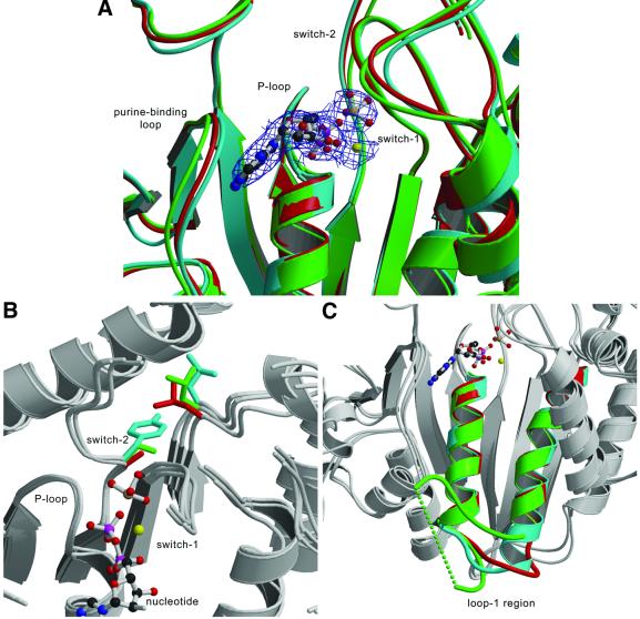
Fig. 2. Comparison of the nucleotide-binding site of MyoE and myosin-II from Dictyostelium and smooth muscle. (A) The nucleotide-binding sites of MyoE (cyan), DdM754 (red) and Gg smII (green) are shown with the MgADP·VO4 from MyoE. (B) Although the sequences of the nucleotide binding regions, including the P-loop, switch-1 and switch-2 are almost entirely conserved, a significant difference occurs in switch-2, where a conserved alanine (red) or serine in amoeboid class-II myosins (green), is replaced by tyrosine (cyan). The introduction of a bulkier side chain pushes a conserved leucine away. (C) Major differences are seen in the length and conformation of loop-1 in MyoE (cyan), DdM754 (red) and Gg smII (green). The green dotted line connects the part of loop-1 that is not visible in the crystal structure of Gg smII.

An official website of the United States government
Here's how you know
Official websites use .gov
A
.gov website belongs to an official
government organization in the United States.
Secure .gov websites use HTTPS
A lock (
) or https:// means you've safely
connected to the .gov website. Share sensitive
information only on official, secure websites.
