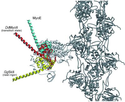
Fig. 4. A model of chicken skeletal muscle myosin motor core (white), converter domain and lever arm (yellow) in the near-rigor state attached to the actin filament (dark gray). Dictyostelium myosin-II in complex with ADP-BeF3 with a modeled extended lever arm in the ‘up’ or transition state position is shown in red. The MyoE converter domain and modeled extended lever arm (cyan) is in an ∼30° higher position.

An official website of the United States government
Here's how you know
Official websites use .gov
A
.gov website belongs to an official
government organization in the United States.
Secure .gov websites use HTTPS
A lock (
) or https:// means you've safely
connected to the .gov website. Share sensitive
information only on official, secure websites.
