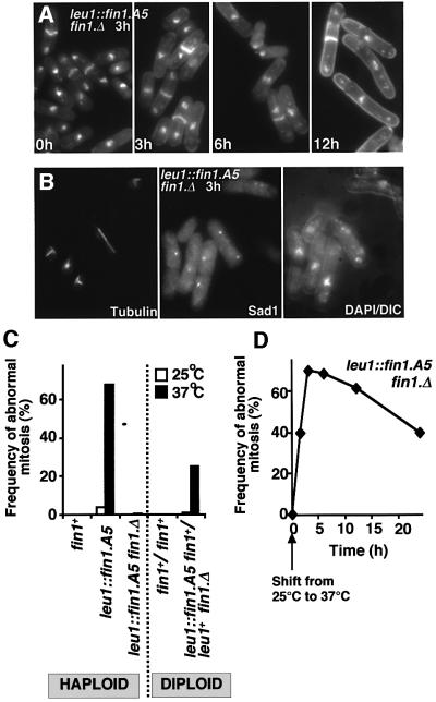
Fig. 2. Temperature-sensitive defects in spindle formation and cell cycle progression in fin1.A5 cells. (A, B and D) fin1.A5 cells were grown to early log phase in EMM2 at 25°C before the temperature of the culture was shifted to 37°C for the times indicated. (A) DAPI/calcofluor staining of fin1.A5 at 37°C. Note the cell elongation 12 h after the temperature shift. (B) Micrographs showing three images of the same field of cells 3 h after the temperature shift. From left to right; microtubules, Sad1 staining and a combined DAPI DIC image. (C) The proportion of mitoses that exhibit spindle formation defects at the indicated temperatures for the haploid and diploid strains is indicated. (D) Mitotic profiles were scored in fin1.A5 cells at distinct time points for 24 h after the temperature shift, and the relative proportion of those that were defective was plotted against time.

An official website of the United States government
Here's how you know
Official websites use .gov
A
.gov website belongs to an official
government organization in the United States.
Secure .gov websites use HTTPS
A lock (
) or https:// means you've safely
connected to the .gov website. Share sensitive
information only on official, secure websites.
