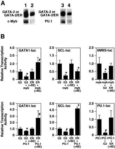
Fig. 7. Differential binding activity and transcriptional activity of GATA-2 and the activated GATA-2/ER. (A) Co-immunoprecipitation analysis of c-Myb and PU.1. 293T cells were transfected with expression plasmids of FLAG-GATA-2 and c-Myb (lane 1), FLAG-GATA-2/ER and c-Myb (lane 2), FLAG-GATA-2 and PU.1 (lane 3) and FLAG-GATA-2/ER and PU.1 (lane 4). β-estradiol was added in the experiments of FLAG-GATA-2/ER (lanes 2 and 4); however, the data were essentially the same in the absence of β-estradiol (data not shown). Total cell lysates were immunoprecipitated with anti-FLAG antibody. The precipitated protein was electrophoresed by SDS–PAGE, blotted and hybridized with the anti-FLAG antibody (upper panels), anti- c-Myb antibody (lower panels of lanes 1 and 2) and anti-PU.1 antibody (lower panels of lanes 3 and 4). (B) Reporter gene analysis of various promoters. Reporter luciferase genes are as described in each panel. G2 and ER stand for the transfection of the GATA-2-expressing plasmid and the GATA-2/ER-expressing plasmid, respectively. When the GATA-2/ER expression plasmid was transfected, β-estradiol was added to activate the protein. An 8 µg aliquot of individual expression plasmids and reporter gene plasmids was transfected into CV-1 cells. Data are shown as the mean ± SD of nine samples (*P < 0.01; #P < 0.05 by t-test).

An official website of the United States government
Here's how you know
Official websites use .gov
A
.gov website belongs to an official
government organization in the United States.
Secure .gov websites use HTTPS
A lock (
) or https:// means you've safely
connected to the .gov website. Share sensitive
information only on official, secure websites.
