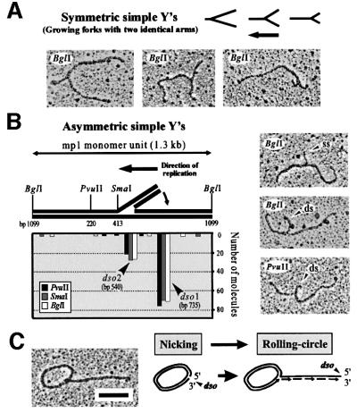
Fig. 2. Mapping of σ-like mp1 replication intermediates with one tail. Plasmid molecules from fractions 2, 3 and 4 were combined and linearized with BglI, PvuI and SmaI, respectively. EM investigation of 100 molecules each revealed symmetric simple Ys (A) and asymmetric simple Ys (B). The location of the origins dso1 and dso2 was deduced as described in the text. The x-axis corresponds to positions on the plasmid map as indicated in the model above. Examples are shown in the right panel. (C) Example and model of an uncut mp1 RC. Bar = 0.5 kb.

An official website of the United States government
Here's how you know
Official websites use .gov
A
.gov website belongs to an official
government organization in the United States.
Secure .gov websites use HTTPS
A lock (
) or https:// means you've safely
connected to the .gov website. Share sensitive
information only on official, secure websites.
