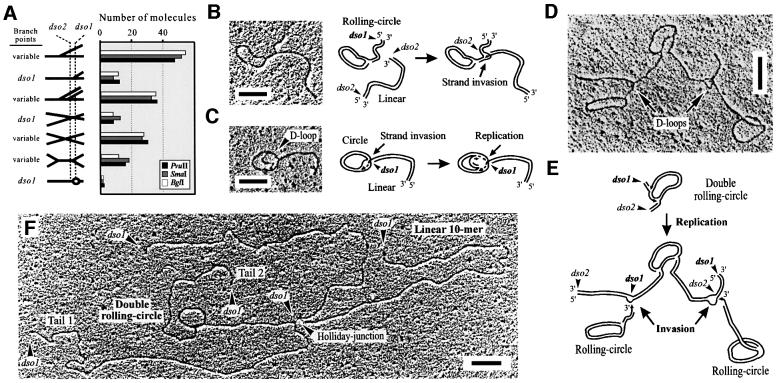
Fig. 7. EM of highly complex in vivo mp1 replication and recombination intermediates. (A) Molecules from fraction 8 were linearized with BglI, PvuI and SmaI, respectively. EM of 150 molecules mainly revealed symmetric simple Ys and double Ys. Examples are shown in the right panel. (B–F) Examples and explanation of uncut complex mp1 concatemers. (B) Open circle with a branched double-stranded tail of 1.3 kb. (C) Open circle with a 1.1 kb tail and a D-loop. (D) Highly branched mp1 molecule (total size 8.9 kb). (E) Model for the origin of this structure. The upper molecule may represent a double RC initiated at dso1 and dso2. Both RC tails are invaded by two other RCs (bottom). (F) Highly branched mp1 molecule (total size is 26.3 kb) that represents a double RC (reinitiated at dso1), recombining with a linear 10mer at dso1. Bars = 0.5 kb.

An official website of the United States government
Here's how you know
Official websites use .gov
A
.gov website belongs to an official
government organization in the United States.
Secure .gov websites use HTTPS
A lock (
) or https:// means you've safely
connected to the .gov website. Share sensitive
information only on official, secure websites.
