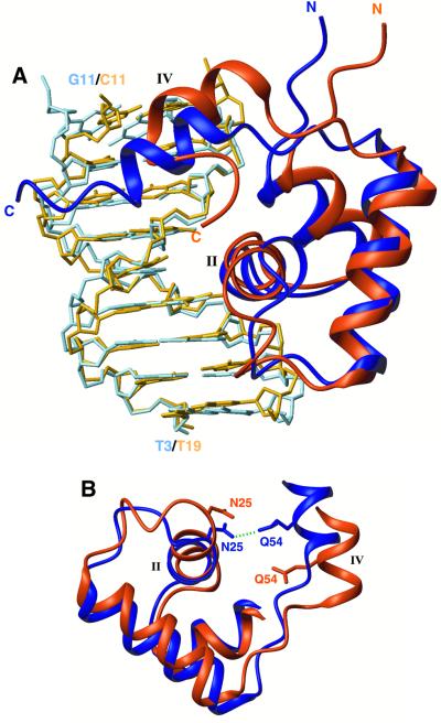
Fig. 3. Superposition of the two O1 operator-bound protein subunits. The left lac headpiece is coloured dark blue and the left half-site of the operator sequence (bp 3–11) is in light blue, whereas the right lac headpiece is coloured dark orange and the right half-site of the operator sequence (bp 11–19) is in light orange. (A) The two structures have been overlaid on their DNA backbone with respect to their approximate dyad-symmetric sequence (i.e. bp 3–11 are overlaid with bp 19–11). The hinge helix in the right site moves by ∼3.4 Å closer to the centre of the sequence and the recognition helix rotates by 12° relative to the left one. (B) In the left site, the hinge helix is stabilized through a hydrogen bond (green dashed line) between Gln54 and Asn25. The extension of the loop linking the third helix with the hinge helix in the right site results in the disruption of this critical contact, thereby diminishing the stability of the right hinge helix.

An official website of the United States government
Here's how you know
Official websites use .gov
A
.gov website belongs to an official
government organization in the United States.
Secure .gov websites use HTTPS
A lock (
) or https:// means you've safely
connected to the .gov website. Share sensitive
information only on official, secure websites.
