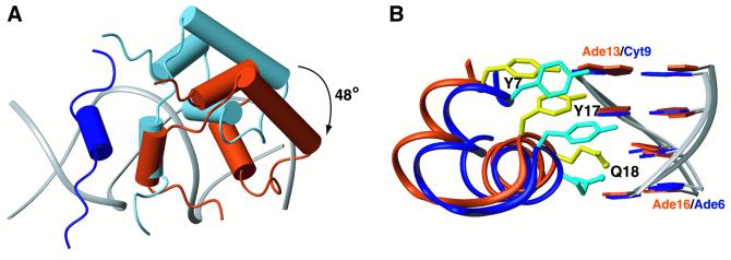
Fig. 6. Distinct recognition mechanisms involved in lac repressor interaction with the left and right operator sites. (A) The left hinge helix is displayed in dark blue. If the lac repressor recognized the right half-site in a similar way to the left site, then it would adopt the conformation displayed in light blue. However, recognition of the right half-site is accomplished by a 48° rotation and a shift of ∼3.4 Å further away from the centre of the operator of the three α-helical domain, whereas the hinge helices still pack together. The calculated conformation of the right subunit of lac repressor’s DBD is displayed in dark orange. (B) Specific recognition of the two sites also requires significant rearrangement of the side chain conformations (the three most important residues, Y7, Y17 and Q18, are depicted) as well as a sequence-dependent deformation of the operator. Dark blue and dark orange denote the left and right site, respectively.

An official website of the United States government
Here's how you know
Official websites use .gov
A
.gov website belongs to an official
government organization in the United States.
Secure .gov websites use HTTPS
A lock (
) or https:// means you've safely
connected to the .gov website. Share sensitive
information only on official, secure websites.
