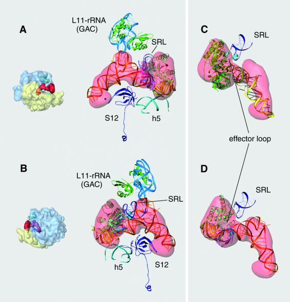
Fig. 4. Interaction of EF-Tu and aa-tRNA with the ribosome. (A and B) Ribbons representation of the docked EF-Tu and aa-tRNA within the ternary complex. (C and D) Focus on the interaction between the α-sarcin–ricin loop (SRL) and the effector loop within domain I of EF-Tu (cyan). In (C) the coordinates of the whole ternary complex from T.aquaticus with a GTP analog (Nissen et al., 1995) were used for the fitting, while in (D) the crystal structure of EF-Tu from E.coli bound to GDP (Song et al., 1999) was used. Orientation of the ribosomes for (A) and (B) are shown as thumbnails on the left. Labeling is the same as in Figure 5.

An official website of the United States government
Here's how you know
Official websites use .gov
A
.gov website belongs to an official
government organization in the United States.
Secure .gov websites use HTTPS
A lock (
) or https:// means you've safely
connected to the .gov website. Share sensitive
information only on official, secure websites.
