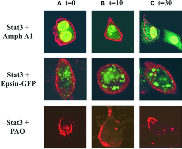
Fig. 7. Inhibition of endocytosis blocks Stat3 translocation to the perinuclear region. Immunofluorescence analysis of NIH-3T3/EGFR cells was carried out after transfection of expression vectors encoding Stat3, Amph A1 or Epsin 2a–GFP. Antibodies to Stat3 or the HA tag of Amph A1 were used to detect localization of these proteins. Transfected cells were treated with 1 µg/ml EGF for 0 min (A), 10 min (B) and 30 min (C). Localization of Stat3 (red) and Amph A1 (green) or Epsin 2a–GFP (green) was analyzed using a Zeiss confocal microscope.

An official website of the United States government
Here's how you know
Official websites use .gov
A
.gov website belongs to an official
government organization in the United States.
Secure .gov websites use HTTPS
A lock (
) or https:// means you've safely
connected to the .gov website. Share sensitive
information only on official, secure websites.
