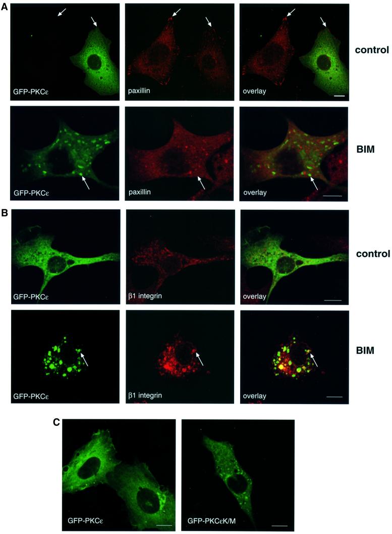
Fig. 3. Cellular localization of GFP–PKCε and paxillin in PKCεKO cells. (A) PKCεKO cells were transiently transfected with GFP–PKCε for 24 h before preparation for microscopy. The cells were plated and allowed to spread on fibronectin for 30 min, followed by a further 90 min incubation either untreated (top) or treated with 1 µM BIM I (bottom). Representative confocal images showing 0.8 µm sections of cells positive or negative for GFP–PKCε also stained with anti-paxillin mAb (red) are shown. The bar represents 10 µm. PKCε decreases the number of prominent focal adhesions detected with paxillin staining (arrows, top) and confirms the accumulation of PKCε in vesicles upon BIM I treatment (arrows, bottom). Note that paxillin is not localized to the GFP–PKCε-positive vesicles but is present in distinct vesicular structures. (B) Integrin β1 co-localizes with GFP–PKCε in vesicles observed in PKCεKO cells upon BIM I treatment. Cells were transfected, plated on fibronectin and either left untreated (top) or treated with BIM I (bottom) as above. Shown are confocal images of cells positive for GFP–PKCε and stained with anti-integrin mAb (red). Examples of sites of co-localization are indicated with arrows. (C) Kinase-dead mutant of GFP–PKCε is expressed in vesicles in untreated cells. PKCεKO cells were transfected with GFP–PKCε or GFP–PKCεK/M for 36 h before preparation for microscopy.

An official website of the United States government
Here's how you know
Official websites use .gov
A
.gov website belongs to an official
government organization in the United States.
Secure .gov websites use HTTPS
A lock (
) or https:// means you've safely
connected to the .gov website. Share sensitive
information only on official, secure websites.
