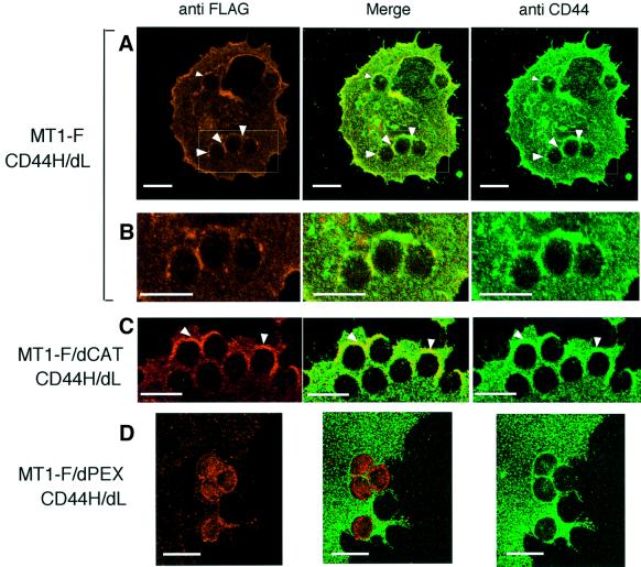
Fig. 5. MT1-MMP movement by pulling CD44H on the cell surface. A Myc-tagged mutant CD44H lacking the ligand-binding domain (CD44H/dL) was expressed together with either MT1-F (A and B), MT1F/dCAT (C) or MT1-F/dPEX (D) in COS-1 cells. After 48 h transfection, cells on glass coverslips were treated with anti-Myc-conjugated microbeads for 30 min. CD44 was then visualized with anti-CD44 rabbit polyclonal antibody and Alexa488-conjugated anti-rabbit IgG antibody. MT1-F, MT1-F/dCAT and MT1-F/dPEX were visualized with anti-FLAG M2 antibody and Alexa568-conjugated anti-mouse IgG antibody. The boxed areas in the MT1-F column (A) are magnified in the lower column (B). Only magnified pictures are presented for MT1-F/dCAT (C) and MT1-F/dPEX (D). Scale bar, 10 µm.

An official website of the United States government
Here's how you know
Official websites use .gov
A
.gov website belongs to an official
government organization in the United States.
Secure .gov websites use HTTPS
A lock (
) or https:// means you've safely
connected to the .gov website. Share sensitive
information only on official, secure websites.
