


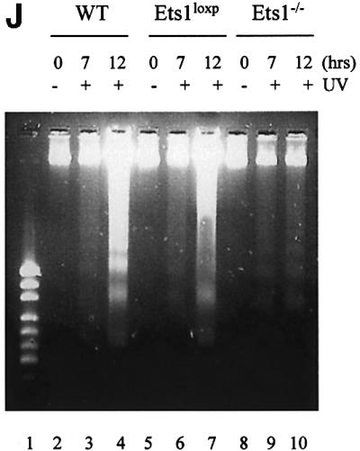
Fig. 3. Ets1–/– ES cells are resistant to UV irradiation-induced apoptosis. (A) Flow cytometric analysis of fixed wild-type, Ets1-targeted and two independent Ets1 null ES cell clones stained with propidium iodide 12 h after UV irradiation (40 J/m2). Percentage of cells with a hypodiploid (sub-2N) DNA content indicative of apoptosis are shown. (B) Graphical representation of the percentage of cells undergoing apoptosis after various doses of UV irradiation. Data is shown as mean ± SD of three independent experiments. (C) Percentage of cells undergoing apoptosis at different time points after irradiation with 40 J/m2 of UV. Data is shown as mean ± SD of three independent experiments. (D–I) Immunofluorescent images of Hoechst 33342-stained WT (D and G), Ets1loxP (E and H) and Ets1–/– (F and I) ES cells, with and without UV irradiation. Significant nuclear condensation and fragmentation is not observed in untreated cells (D–F) or UV-irradiated Ets1 null cells (I). However, a significant number of WT and Ets1-targeted ES cells showed nuclear fragmentation after UV irradiation (G and H). Bar corresponds to 20 µm. (J) Ethidium bromide staining of DNA from WT, Ets1loxP and Ets1–/– ES cells after 40 J/m2 UV irradiation analysed by agarose gel electrophoresis, demonstrating bands of DNA fragmentation in WT and targeted but not Ets1–/– cells. Lane 1 shows pUC19/HpaII marker.