

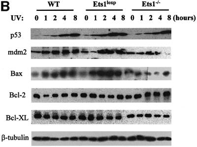
Fig. 5. The expression of p53 transactivated genes is reduced in cells lacking Ets1 following UV irradiation. (A) Results of a typical northern blot experiment measuring the expression of p53-regulated genes in wild-type (wt), Ets1-targeted (Ets1loxP) and Ets1–/– ES cells before and after UV irradiation (40 J/m2). Cells were analysed and RNA isolated at times indicated after UV treatment. Probes specific for mouse perp, mdm2, cyclin G and bax were used for northern blot analyses with GAPDH as a loading control. Data from five independent experiments of two independent Ets1–/– clones were quantified using a Fuji Image Reader VI.3E and expressed as mean ± SD, relative to a GAPDH loading control. (B) Expression of p53-regulated proteins in wild-type and Ets1–/– ES cells. Cells were isolated at the indicated time points (0, 1, 2, 4 and 8 h) post-UV irradiation (40 J/m2). Antibodies specific for mouse p53, mdm2, Bax, Bcl-2, Bcl-XL and β-tubulin (loading control) were used for western blot analyses. Analysis was performed at least three times on two independent Ets1–/– clones and data from a representative experiment is shown.