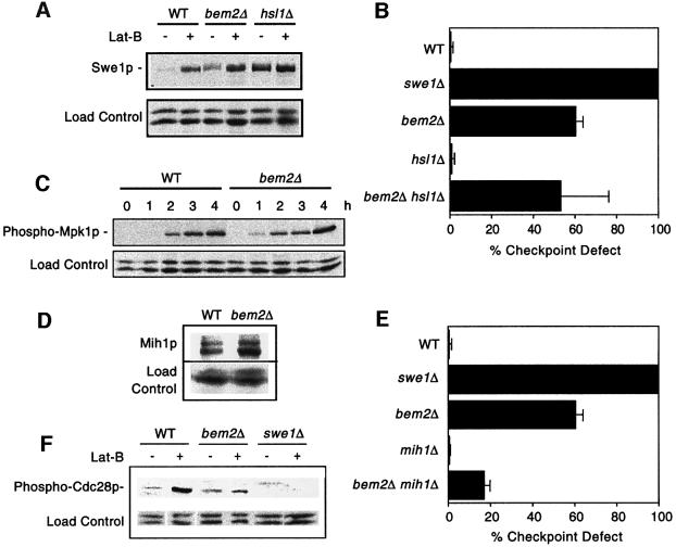
Fig. 7. Swe1p, Mpk1p and Cdc28p regulation in bem2Δ cells. (A) The strains DLY1 (WT), DLY4021 (swe1Δ), DLY4015 (bem2Δ), JMY3-1 (hsl1Δ) and DLY4019 (bem2Δ hsl1Δ) were assayed for checkpoint function as in Figure 2A. (B) The strains DLY5330 (SWE1-myc), DLY5331 (bem2Δ SWE1-myc) and JMY1503 (hsl1Δ SWE1-myc) were grown to exponential phase and then resuspended in 100 µM Lat-B in YEPD and cultured for 2 h at 30°C. Lysates from cells before and after growth in Lat-B were immunoblotted with α-myc antibody. (C) The strains DLY1 (WT) and DLY4015 (bem2Δ) were grown to exponential phase in YEPD + 0.4 M NaCl, followed by resuspension and growth for the indicated times in the same medium supplemented with 100 µM Lat-B. Lysates were immunoblotted with anti-phospho-p44/p42 MAPK antibody to detect activated Mpk1p. (D) The strains JMY1299 (MIH1-myc) and DLY 4478 (bem2Δ MIH1-myc) were grown to exponential phase. Lysates were immunoblotted with α-myc antibody. (E) The strains DLY1 (WT), DLY4021 (swe1Δ), DLY4015 (bem2Δ), JMY3-59 (mih1Δ) and DLY3860 (bem2Δ mih1Δ) were assayed for checkpoint function as in Figure 2A. (F) The strains DLY1 (WT), DLY4015 (bem2Δ) and DLY4021 (swe1Δ) were grown to exponential phase in YEPD at 24°C and then resuspended in 100 µM Lat-B in YEPD and cultured for an additional 2 h. Lysates from cells before and after growth in Lat-B were immunoblotted with an anti-phospho-cdc2 antibody to visualize Tyr19 phosphorylated Cdc28p.

An official website of the United States government
Here's how you know
Official websites use .gov
A
.gov website belongs to an official
government organization in the United States.
Secure .gov websites use HTTPS
A lock (
) or https:// means you've safely
connected to the .gov website. Share sensitive
information only on official, secure websites.
