Abstract
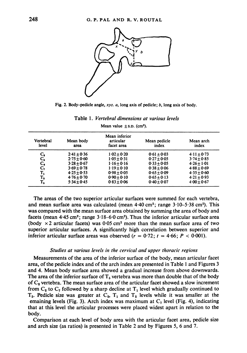
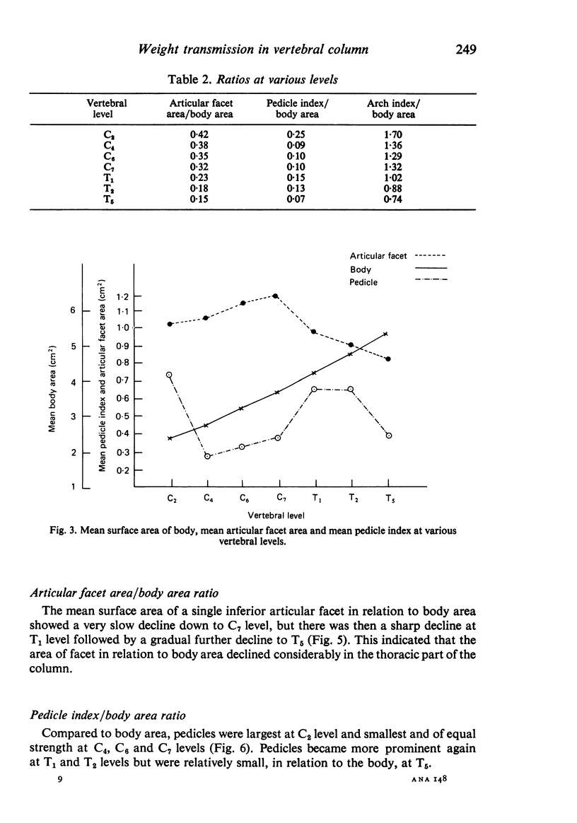
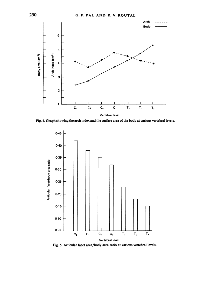
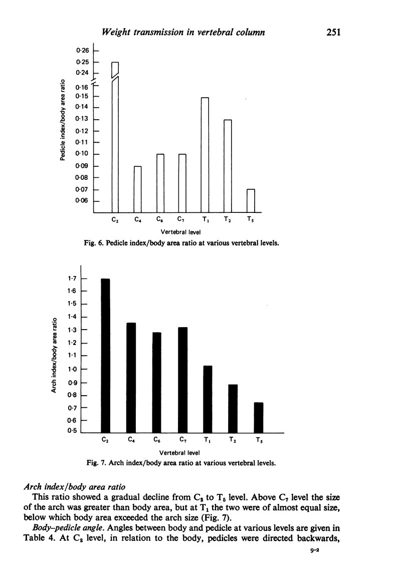
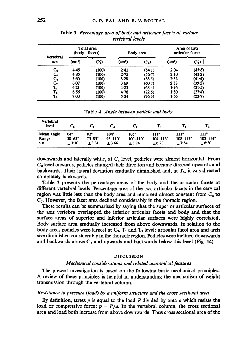
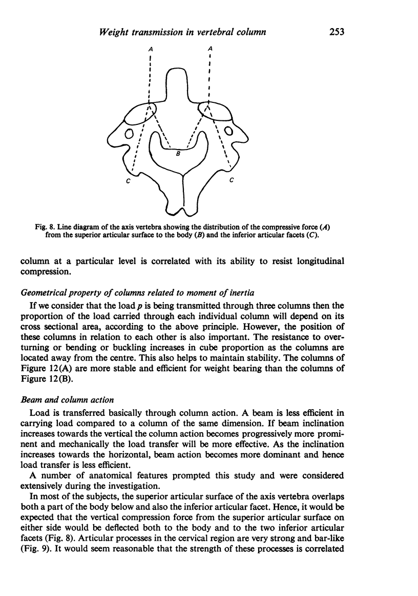
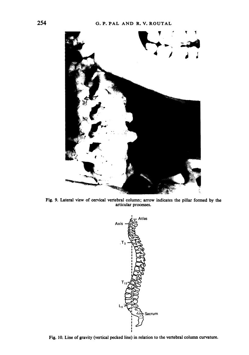
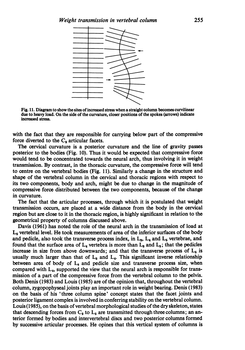
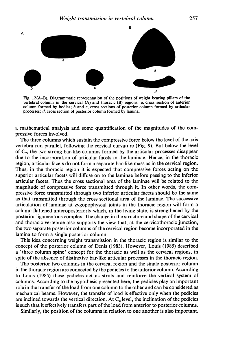
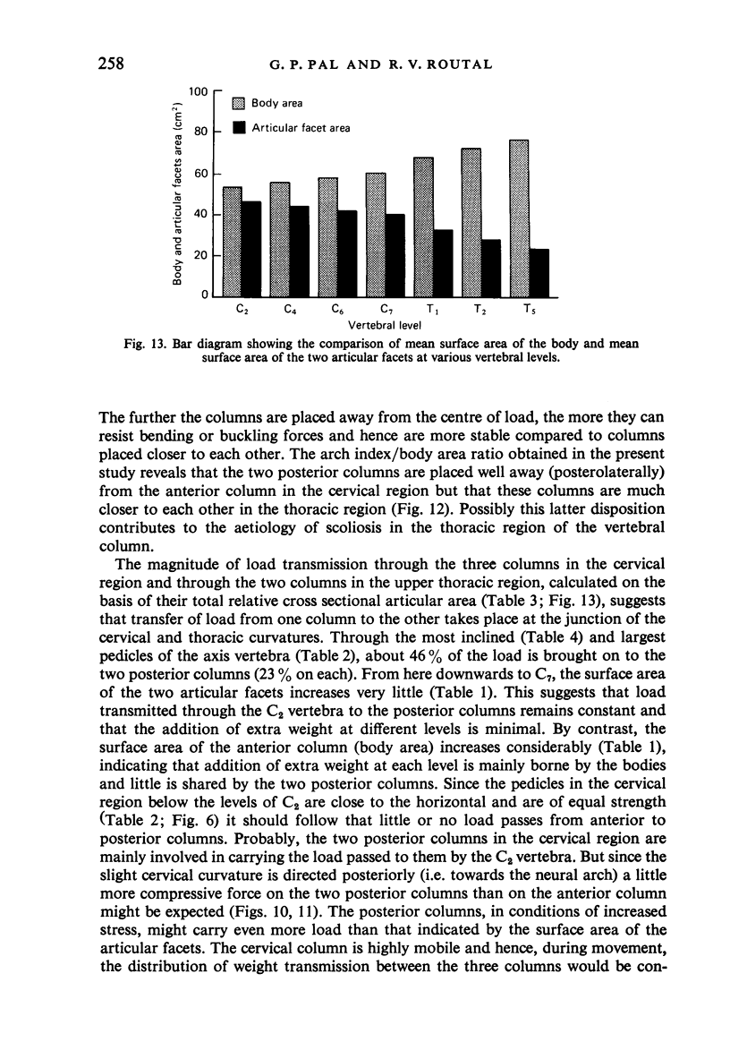
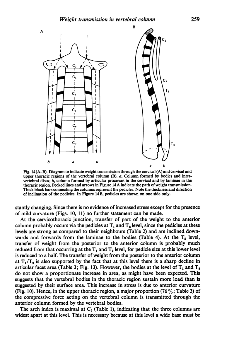
The role of the neural arch in weight transmission in the cervical and upper thoracic regions of the vertebral column has been investigated. Measurements at the levels of C2, C4, C6, C7, T1 and T5 vertebrae were made in 44 adult male vertebral columns. At each level, the area of the inferior surface of the body was compared with the area of the inferior articular facets, the pedicle index and the arch index; inclination of the pedicle in relation to the body was also measured. On the basis of these studies it was found that at C2 level the compressive force acting on the superior articular surfaces was transmitted to the inferior surface of the body and to the two inferior articular facets. From C2 to C7, compressive force is transmitted through three parallel columns - one anterior, formed by the bodies and intervertebral discs, and two posterior, formed by the articulations of the articular processes on either side. Due to the posterior curvature in the cervical region, the posterior columns here sustain more of the compressive force. From C7 level downwards, the compressive force is transmitted through two columns, i.e. one anterior formed by the bodies and intervertebral discs and one posterior formed by successive articulations of the laminae. Below C7 level, compressive force from the posterior column is partly transferred to the anterior column through the pedicles at T1 and T2. In the upper thoracic region, due to the anterior curvature, the main part of the compressive force is transmitted through the anterior column, which sustains even greater compressive force than is suggested by body area, with resulting increased stress.
Full text
PDF
Images in this article
Selected References
These references are in PubMed. This may not be the complete list of references from this article.
- Brandner M. E. Normal values of the vertebral body and intervertebral disk index during growth. Am J Roentgenol Radium Ther Nucl Med. 1970 Nov;110(3):618–627. doi: 10.2214/ajr.110.3.618. [DOI] [PubMed] [Google Scholar]
- DAVIS P. R. The medial inclination of the human thoracic intervertebral articular facets. J Anat. 1959 Jan;93(1):68–74. [PMC free article] [PubMed] [Google Scholar]
- Davis P. R. Human lower lumbar vertebrae: Some mechanical and osteological considerations. J Anat. 1961 Jul;95(Pt 3):337–344. [PMC free article] [PubMed] [Google Scholar]
- Denis F. The three column spine and its significance in the classification of acute thoracolumbar spinal injuries. Spine (Phila Pa 1976) 1983 Nov-Dec;8(8):817–831. doi: 10.1097/00007632-198311000-00003. [DOI] [PubMed] [Google Scholar]
- Taylor J. R., Twomey L. T. Sexual dimorphism in human vertebral body shape. J Anat. 1984 Mar;138(Pt 2):281–286. [PMC free article] [PubMed] [Google Scholar]


