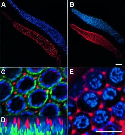
Fig. 2. LIP-1 expression and localization in the distal gonad arm. LIP-1 antibody staining (in red) and DAPI staining (in blue) in a dissected distal gonad arm of (A) a 2- to 3-day-old wild-type adult hermaphrodite and (B) a lip-1(0) mutant of similar age. (C) Higher magnification view of pachytene germ cells in a wild-type gonad stained with the LIP-1 antibody (in red), the nuclear pore complex antibody mAB414 (Yamada and Kasamatsu, 1993) (in green) and DAPI (in blue). (D) An xz-view of the same region as shown in (C). LIP-1 staining was concentrated in ∼2 µm long rod-like structures originating in the outer region near the pachytene nuclei and facing towards the cytoplasmic core. (E) Co-staining of LIP-1 with the hydrophobic dye Dil that labels the plasma membrane surrounding the pachytene germ cells. LIP-1 was concentrated at the junctions where membranes surrounding individual cells are connected. (A–C) Optical sections through the outer region of the distal syncytial gonad containing the germ cell nuclei. (D and E) Three-dimensional reconstructions of the entire data stacks (for details see Materials and methods). The scale bar in (B) is 20 µm and in (E) 5 µm.

An official website of the United States government
Here's how you know
Official websites use .gov
A
.gov website belongs to an official
government organization in the United States.
Secure .gov websites use HTTPS
A lock (
) or https:// means you've safely
connected to the .gov website. Share sensitive
information only on official, secure websites.
