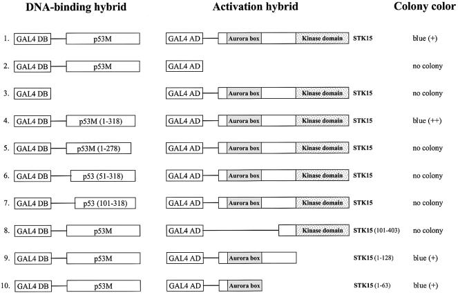
Fig. 1. Characterization of the p53–STK15 interaction in yeast. Left column: GAL4 DNA-binding domain (amino acids 1–147) hybrids. Middle column: GAL4 activation domain (amino acids 768–881) hybrids. The STK15 fragment fused to GAL4 activation domain is indicated on the right of the diagrams. The relative positions of the Aurora box and kinase domains of STK15 are also depicted. Right column: yeast colony color after transformation; the relative color is in parentheses. No colony indicates that no yeast colony was observed after incubation for a standard period of time.

An official website of the United States government
Here's how you know
Official websites use .gov
A
.gov website belongs to an official
government organization in the United States.
Secure .gov websites use HTTPS
A lock (
) or https:// means you've safely
connected to the .gov website. Share sensitive
information only on official, secure websites.
