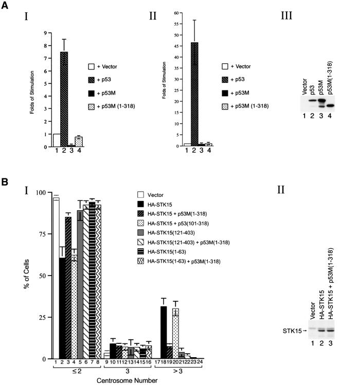
Fig. 4. Blocking of STK15-mediated centrosome amplification by a transactivation-defective p53 mutant. (A) Transactivation assay. Panel I: a luciferase reporter driven by three p53-binding sites was co-transfected with vector alone (lane 1), p53 (lane 2), p53M (lane 3) or p53M(1–318) (lane 4). Panel II: as in panel I, except that the reporter was driven by the p21 promoter. Panel III: protein expression levels of p53 derivatives. Lysates from NIH-3T3 cells transfected with vector alone (lane 1), p53 (lane 2), p53M (lane 3) or p53M(1–318) (lane 4) were blotted with an anti-p53 antibody. (B) Inhibition of STK15-mediated centrosome amplification by p53M(1–318). Panel I: centrosome numbers in 900 NIH-3T3 cells transfected with the specified plasmids were counted by immunostaining with an anti-γ-tubulin monoclonal antibody. The combination of plasmids transfected is indicated. Experiments were repeated three times. Panel II: protein expression levels of STK15. Lysates from NIH-3T3 cells transfected with vector alone (lane 1), HA-STK15 or HA-STK15 plus p53M(1–318) were blotted with an anti-HA antibody.

An official website of the United States government
Here's how you know
Official websites use .gov
A
.gov website belongs to an official
government organization in the United States.
Secure .gov websites use HTTPS
A lock (
) or https:// means you've safely
connected to the .gov website. Share sensitive
information only on official, secure websites.
