

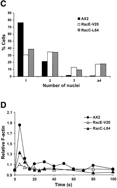
Fig. 8. Expression of constitutively active Rho GTPases RacC-L64 and RacE-V20 fused to GFP partially phenocopy the defects of GDI– cells. (A) Growth in shaking suspension. Curves are representative of two independent determinations, each performed in duplicate. (B) Nuclear staining and actin distribution. From top to bottom, phase–contrast, DAPI staining and immunostaining of actin of cells grown in suspension. Cells were allowed to sit for 20 min on coverslips prior to fixation. Scale bar, 25 µm. (C) Distribution of the number of nuclei in mutants grown in suspension. (D) Actin polymerization upon cAMP stimulation of aggregation competent cells. Experiments were performed as described in Figure 6A. Both mutants display growth, cytokinesis and actin polymerization defects.