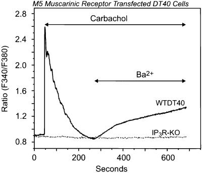
Fig. 3. Activation of the G-protein-coupled M5 muscarinic receptor results in stimulation of Ba2+ entry in wild-type but not in IP3R-KO DT40 cells. Ba2+ influx was measured in single Fura-2-loaded wild-type (WTDT40) or IP3R-KO (dotted line) DT40 cells transfected with the human M5 muscarinic receptor. The cells were maintained in nominally Ca2+-free medium, exposed to 100 µM carbachol, and then Ba2+ (10 mM) was added where indicated. The relatively higher ratio values in this experiment are due to the use of a different Ca2+ measuring system; in this series, a photon counting system was used, while in the other series, an imaging system was used. Representative traces from three independent experiments are shown.

An official website of the United States government
Here's how you know
Official websites use .gov
A
.gov website belongs to an official
government organization in the United States.
Secure .gov websites use HTTPS
A lock (
) or https:// means you've safely
connected to the .gov website. Share sensitive
information only on official, secure websites.
