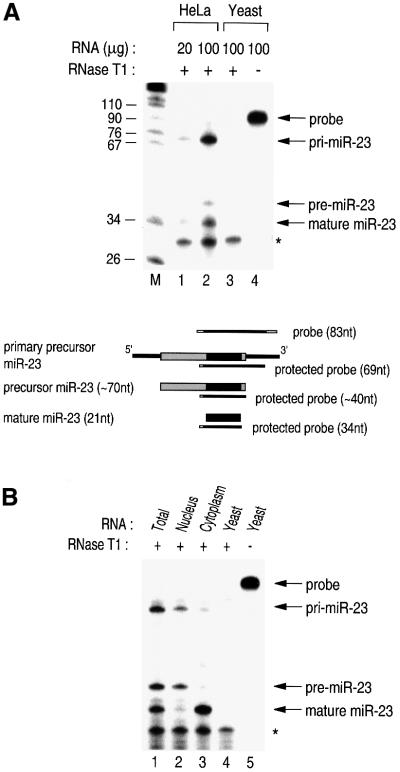
Fig. 5. Subcellular localization of the three different forms of miRNA. (A) RPA to detect miRNA. The indicated amounts of RNA from either HeLa or yeast cells were used for RPA. The asterisk indicates the self-protected probe. Schematically represented are the probes for the RPA and the protected fragments that are expected for each miRNA species. (B) Total, nuclear or cytoplasmic RNA from HeLa cells or yeast RNA was used for RPA.

An official website of the United States government
Here's how you know
Official websites use .gov
A
.gov website belongs to an official
government organization in the United States.
Secure .gov websites use HTTPS
A lock (
) or https:// means you've safely
connected to the .gov website. Share sensitive
information only on official, secure websites.
