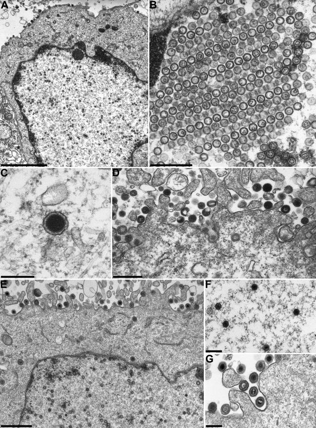
FIG.8.
Electron microscopy of PrV-ΔUL17F-infected cells. RK13 (A to D) and RK13-UL17 (E to G) cells were infected with PrV-ΔUL17F at an MOI of 1 and processed for electron microscopy 14 h p.i. (A) Overview of an infected RK13 cell. (B) Pseudocrystalline aggregations of B-capsids in the nucleus. (C and D) L-particles in the cytoplasm (C) and on the cell surface (D). (E) Unimpaired virus morphogenesis on UL17-expressing cells, including production of C-capsids in the nucleus (F) and mature virions on the cell surface (G). Bars represent 2 μm in panel A, 500 nm in panels B and D, 250 nm in panels C, F, and G, and 1 μm in panel E.

