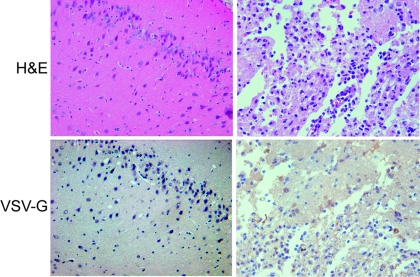
FIG. 6.
Representative photomicrographs of rat brains from animals at day 14 after injection with 4 × 10⁷ PFU of rVSV-F with (left) and without (right) prophylactic rat IFN-α treatment. Top panels, H&E staining; bottom panels, VSV-G immunohistochemical staining; magnification, ×40. The left panels show intact brain tissue without evidence of virus-induced necrosis. The right panels show a region of virus-induced necrosis with positive immunohistochemical staining for VSV-G in several cells within the necrotic focus.

