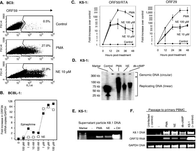
FIG. 1.
Effect of norepinephrine (NE) on KSHV reactivation from latency. (A) Expression of the KSHV lytic cycle protein ORF59 was assayed by flow cytometry using indirect immunofluorescence in BC3 cells fixed and permeabilized 24 h after exposure to 20 ng/ml PMA, 10 μM norepinephrine, or an equivalent volume of vehicle. (B) Dose dependence of catecholamine effects on KSHV lytic gene expression were also assessed by quantitative real-time RT-PCR detection of KSHV ORF29 mRNA expression (normalized to GAPDH mRNA) 24 h following exposure of BCBL-1 cells to indicated concentrations of epinephrine or norepinephrine. Similar results were observed for ORF50/RTA mRNA (data not shown). (C) Kinetics of norepinephrine-induced lytic gene expression were assessed for immediate-early ORF50/RTA and late lytic ORF29 at the indicated time points by real-time RT-PCR (data represent mean ± standard error fold change in target mRNA concentration in three replicates after normalization to cellular GAPDH). Norepinephrine induction of ORF50/RTA mRNA was statistically significant at all time points (12 h, P < 0.0001; 24 h, P = 0.0011; 36 h, P = 0.0004; 48 h, P = 0.0027; all by t test on log-transformed induction values). Norepinephrine induction of ORF29 was not significant by 12 h (P = 0.2086), but was highly significant at all subsequent time points (24 h, P = 0.0003; 36 h, P = 0.0006; 48 h, P = 0.0003). Similar kinetics were observed in BCBL-1 cells (data not shown). (D) Gardella gel analysis of replicating KSHV DNA was carried out by electrophoresis of KS-1 cell-associated DNA and hybridization with a 32P-labeled PCR amplicon from the KSHV ORF50 locus to distinguish episomal genomic DNA from linear replicating DNA. Data show cells treated for 48 h with vehicle (Control), PMA, norepinephrine, or the PKA activator db-cAMP. (E) KSHV particle-associated DNA was assayed by PCR amplification of KSHV K8.1 sequences in 20 μl of DNase-treated 0.45-μm-filtered supernatant from KS-1 cell cultures treated with vehicle, PMA, or norepinephrine for 48 h (positive control is KS-1 cell-associated DNA). (F) Presence of infectious KSHV in cell culture supernatants from E was assessed by incubating PHA-stimulated PBMC in filtered/DNased supernatants for 1 h followed by washing. 24 h later, PBMC were assayed for KSHV K8.1 DNA and cellular GAPDH by real-time PCR. PBMC not exposed to PEL cell supernatants served as negative controls, and KS-1 cells served as positive controls.

