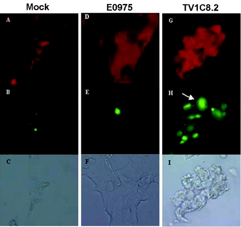
FIG. 2.
Fusogenicity of gp160. Red fluorescence (A, D, and G), green fluorescence (B, E, and H), and bright-field (C, F, and I) images were obtained 8 h after coculturing PM-1 cells with 293T cells expressing no env gene (mock) or the env genes derived from HIV-1-CAR402 or HIV-1 subtype C TV1c8.2. For subtype C gp160TV1c8.2 (G to I), the green fluorescence observed in lymphocytes spreads, forming larger areas of diffuse green fluorescence. The arrowhead indicates an example of colocalization of green and red fluorescence. Controls were PM-1 cells incubated with mock-transfected 293T cells (A to C) or PM-1 cells cultured with consensus-subtype C envelope-E0975-transfected 293T cells (D to F). In these control cocultures, the CMFDA-labeled lymphocytes maintained a uniform, rounded morphology and showed little adherence to the 293T cells. Hardware: Macintosh G4; software: Adobe Photoshop 7.0.

