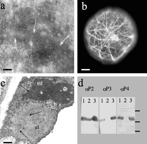
FIG. 1.
Characterization of biochemical and biological properties of mutant P2Rev5. (a) Crude extracts of Sf9 insect cells infected with a baculovirus recombinant expressing P2Rev5, observed by negative staining and electron microscopy. White arrows indicate P2Rev5 paracrystal bundles. (b) Live Sf9 insect cell infected with a baculovirus recombinant expressing a P2Rev5-GFP fusion observed by epifluorescence microscopy. (c) Plant cell infected with CaMV Top-S-Rev5. The cell contains both electron-dense (ed) and electron-lucent (el) inclusion bodies; virions are indicated by black arrows. (d) Far-Western experiments revealing P2-P3 interaction. Ten micrograms of wild-type P2, P2157m (a negative control that can no longer bind P3 [21, 37]), and P2Rev5 were loaded in lanes 1, 2, and 3, respectively. The proteins are specifically revealed with an anti-P2 serum (3) in the left panel and tested for P3 and P3-virion binding capacity in the middle and right panels, respectively (see Materials and Methods). Molecular mass marker positions 6.5, 16.5, and 25 kDa are shown on the right. Bars represent 100, 1,000, and 500 μm in panels a, b, and c, respectively.

