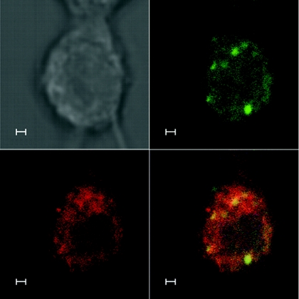
FIG. 3.
Partial colocalization of LMP1 and galectin 9 in NPC cells. C15 NPC cells were double stained with a mouse anti-LMP1 (OT22CN; red secondary antibody, Alexa 546) (lower left) and a rabbit anti-Galectin 9 (green secondary antibody, Alexa 488) (upper right). Overlay (lower right). Scale bar, 1 μm. In this typical C15 cell, galectin 9 and LMP1 colocalize in dots where galectin 9 staining is prominent.

