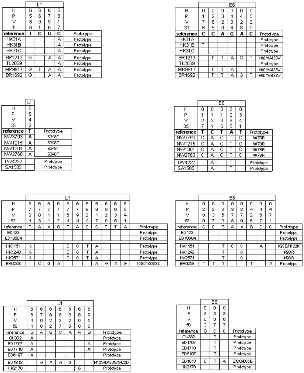
FIG. 7.
Diversity of the E6 genes (four panels on the right side of the figure) and part of the L1 genes (left side of the figure) in distantly related variants of HPV-31, -35, -52, and -58. Within each panel, the first column lists the variants, whose relative phylogenetic position can be found in Fig. 1, 3, 5, and 6. The central part of the figure identifies nucleotide exchanges (letters) or maintenance of the sequence of the reference genome (gray squares). The box on the right side of each panel indicates whether the amino acid sequence of the reference clone has been maintained (“prototype”) and if not, what kind of amino acid exchanges have occurred.

