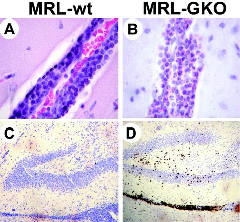
FIG. 6.
BDV infection induces influx of eosinophilic granulocytes into brains of old MRL-GKO mice but not of wild-type mice. MRL wild-type and MRL-GKO mice infected at ages of 60 to 130 days with BDV were sacrificed at the peaks of neurological disease at days 25 to 30 p.i. Paraffin sections of brains were either stained with hematoxylin/eosin (A and B) or for cyanide-resistant peroxidase activity by brown stain which specifically labels eosinophilic granulocytes (C and D). Representative infiltrates around blood vessels in the hippocampus (panels A and B) and dentate gyrus regions at lower magnification (panels C and D) are shown.

